Introduction
Digital technology has revolutionized the way dentists and prosthetists can treat their patients. Digital tools make 2D, 3D, and even 4D (with jaw motion and motion capture solutions) data collection possible, allowing us to optimize our treatments while meeting the patient’s demand for rapid, predictable results. To meet these expectations we must create reliable, synergistic protocols involving both the dental practice and the laboratory, and this is fortunately what digital tools allow us to do today.
This case uses a combination of over 10 different digital tools: a synergy of devices and software from different companies all working and integrating together to enable digital dentistry and ultimately deliver a high-quality long-term full arch prosthesis that improves the quality of life of the patient in terms of both function and aesthetics.
Presentation of the clinical case
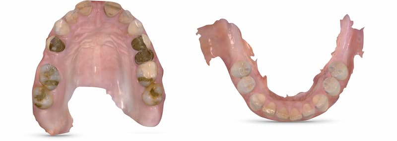
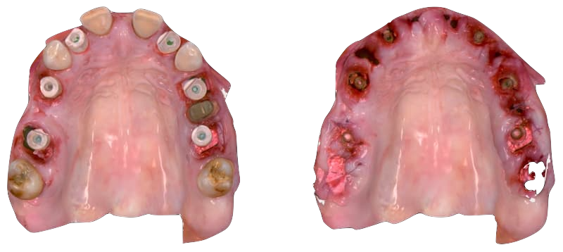
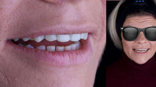
A 47-year-old female patient was referred by her periodontist for an implant treatment. Exhausted by a palliative periodontal maintenance for many years, she could no longer tolerate the dental mobility, the slippage, the decrease in vertical dimension and the spaces that have appeared between her teeth.
The patient wanted a complete fixed implant-supported rehabilitation.
Collection of Digital Data
For these treatments, we have established a strict protocol of digital data collection that aims to create a digital avatar of our patient and also to generate a virtual treatment plan prior to any intervention. This is the basis for the presentation to the patient as well as for preparing an estimate for the treatment.
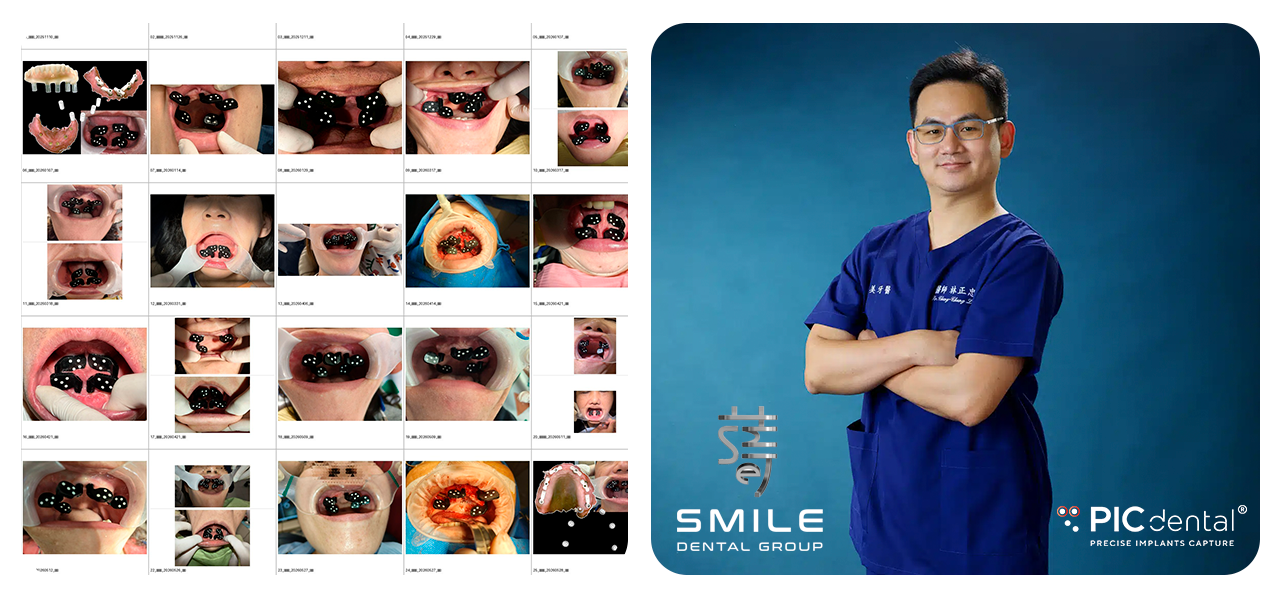
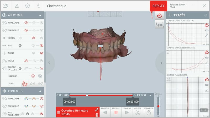
The first appointment is a crucial moment of interaction with the patient to understand her expectations and also to collect all the digital information of her patients records. The collected records in this case included photography, intraoral scanning, motion capture, facial scanning and CBCT.

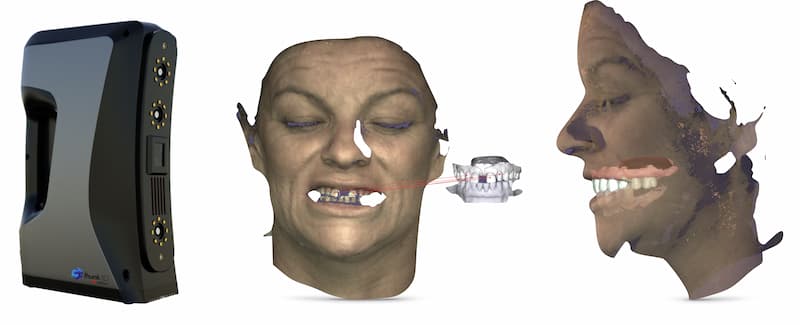
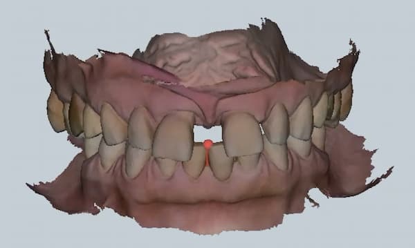
All these elements combined in a smile design software allow us to work with precision on a virtual replica of the patient. With dental morphologies, occlusal plane, position of free edges, occlusion, bone volumes, implant positions... the possibilities when reviewing the case are wide and allow a global approach.
Data Processing - Synergy with the Prosthesis Laboratory
This virtual clone allows the dentist and the dental technician to plan the prosthetic rehabilitation. It will be used to guide the surgical choices and, in particular, the implant placement.
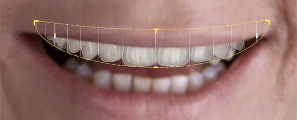
In this specific clinical case, the Smile Cloud® 2D planning made it possible to define a reconstruction space, a position of the free edges, a morphology and to decide on a rehabilitation that follows natural dental emergence profiles. The 3D dental library available for exocad® was downloaded. The facial scan was merged with the intraoral scan, the CBCT and the motion capture of occlusal temporality recorded with MODJAW®.
By this point, the dental technician has a full digital copy of the patient and can work on the different three-dimensional layers.
Taking into account all of these elements, we decided to recenter and raise the vertical dimension of the patient using the rotational movement isolated during the search for an occlusal plane. The system then transposes all the recorded movements to this new vertical dimension and we proceeded with the design stage of the prosthetic project in exocad® with this new vertical dimension. The dental morphologies used are those from the Smile Cloud®. Once the final project is validated, the surgical execution of the case is prepared.
Surgery
Based on current scientific literature, we used static tooth-supported surgical guides in order to maximize precision and sequential extractions (preserving teeth during surgery) in order to improve the quality of intraoral scanning by providing better landmarks.
Two sets of files were prepared for implant planning using 3Shape Implant Studio® software, after which the surgical guides were designed and 3D printed with the SprintRay® printing process. The resins and the post-processing used ensure the biomedical certification of the custom-printed guide.
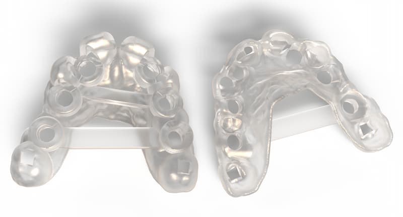
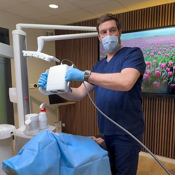
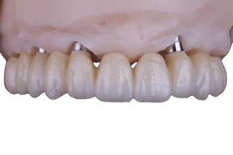
On the day of surgery, after the placement of implants using the surgical guide, intraoral scan data was used to design (exocad®) and print (SprintRay®) a transitional bridge in C&B MFH resin that was placed directly on the multi-unit abutments.
A post-surgery check-up appointment was scheduled on day 10 when a milled duplicate of the transitional bridge made of MultiStratum long-lasting PMMA was placed.
Final prosthetic restoration
Using the PIC system (precise implants capture) for a guaranteed passive fit
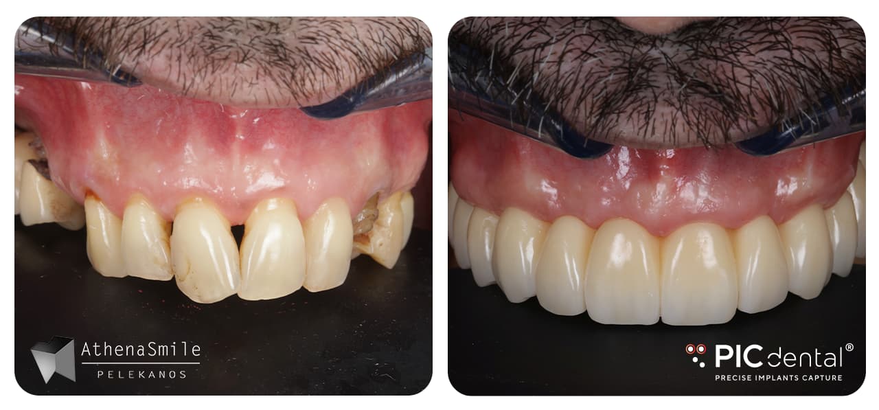
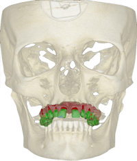
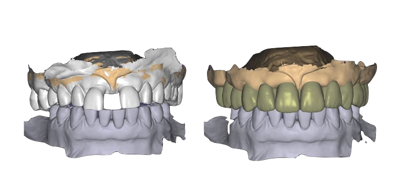
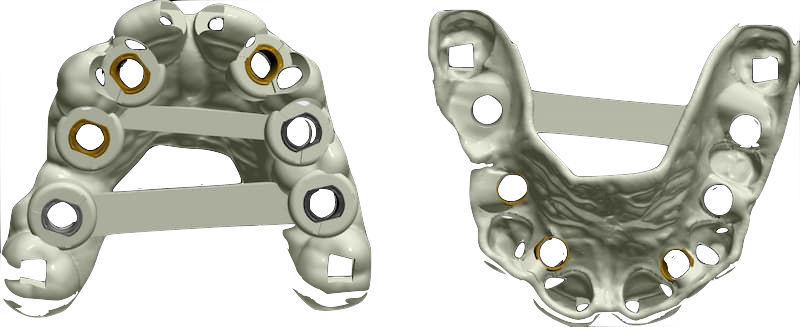
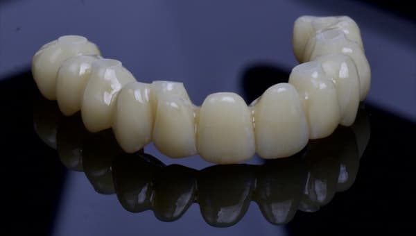
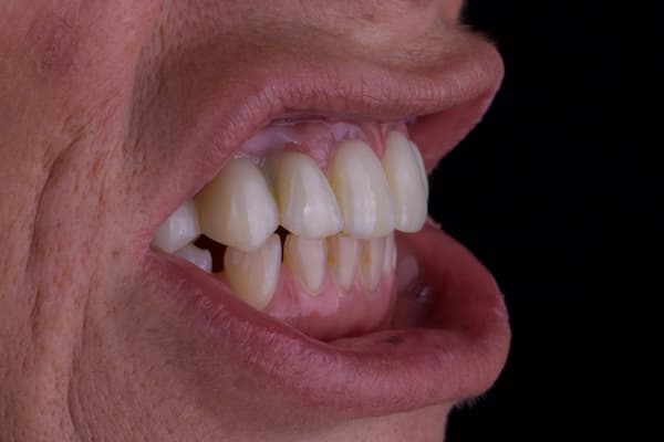
At 3 months post-surgery, our goal was to produce a full zirconia bridge with the emergence profile, without ti-bases, placed directly on the multi-unit abutments.
The known limitation of intraoral scanners is their relative inaccuracy for full arch implant restorations. It has been demonstrated that the predictability of an intraoral scan on 4 or 6 implants is directly dependent on the operator’s experience and technique. While the inaccuracy of some intraoral scanners may be acceptable today when producing machined titanium bars topped with resin teeth, it is unfortunately not sufficient for full zirconia restorations.
![]()
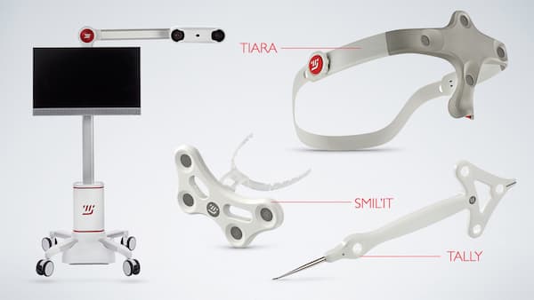
Therefore, the PIC system was used to complete the final prosthetic restoration.
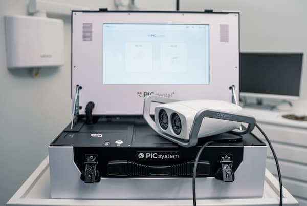 The PIC system is an extremely fast and precise tool for measuring implant positions using photogrammetry.
It is precise up to 4 microns and provides predictable results that are not affected by the operator's technique and experience. It’s the only digital impression technique for measuring multiple implants that provides a guaranteed passive fit validated by over 19 peer-reviewed scientific studies since its initial release in 2010.
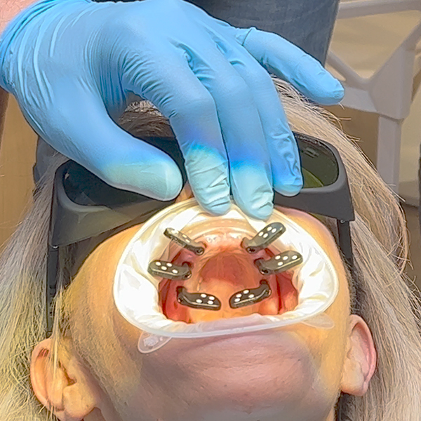
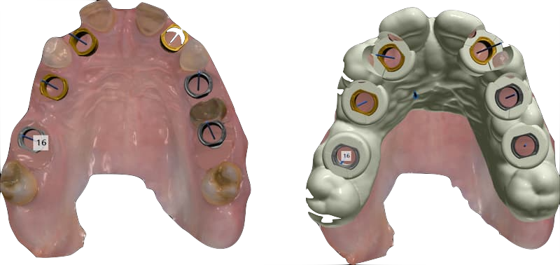
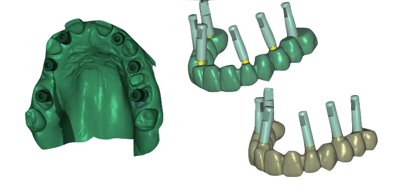
The PIC system generates a PIC file that contains only the interrelated implant positions and angulations. This PIC file is exported in the open STL format that can be used directly in exocad®.
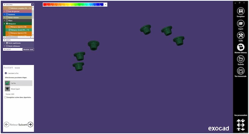
The PIC file was imported into our exocad® prosthetic project, along with an intraoral scan of the healed soft tissues.
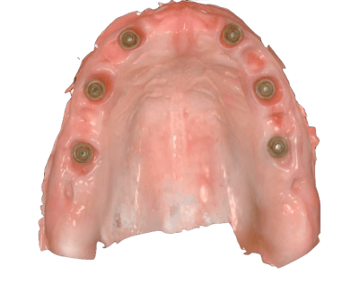
The bridge was designed using the implant measurements from the PIC file, then machined in zirconia YML. The connection is made directly on the multi-unit abutments, without any ti-bases. The advantage of using a zirconia such as the KATANA™ YML is its duality of gradient, both mechanical and esthetic with a variation in translucency.
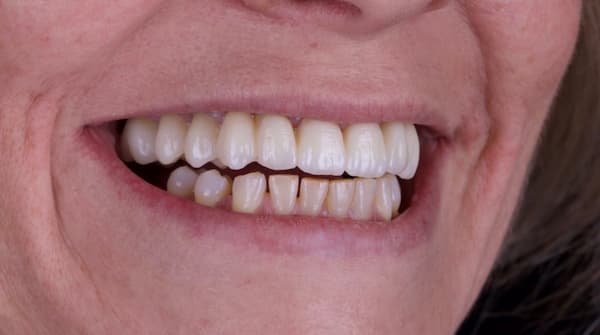
The bridge was placed with a completely passive fit and then tightened with a torque wrench.
Summary of tools used
Appointment 1 (Patient records)
- 2D data collection
- DSLR camera
- Smile Cloud® software for smile design
- 3D data collection
- Intraoral scanner (3Shape® TRIOS)
- Motion capture (MODJAW®)
- Facial scanner (Thunk3D® Archer S)
- CBCT
Prosthetic rehabilitation planning
- Prosthesis design (exocad®)
- Implant planning (3Shape Implant Studio® software)
- 3D printed surgical guides (SprintRay®)
Appointment 2 (Surgery)
- 3D printed transitional bridge (SprintRay®)
-
Intraoral scanner (3Shape® TRIOS)
Appointment 3 (10 days post-surgery check-up)
- Milled provisional PMMA bridge (imes-icore®)
Appointment 4 (3 months post-surgery check-up)
- Precise implants capture (PIC dental® PIC system)
- Intraoral scanner (3Shape® TRIOS)
Appointment 5 (Final restoration)
- Milled full zirconia bridge (imes-icore®)
Conclusion
This simple and repeatable protocol allows the creation of a virtual avatar of the patient by means of 2D, 3D, and 4D data collection. This virtual patient remains consistent throughout the treatment while working on the prosthetic restoration and the surgical planning, with the only additional layers being the information of the implant positions and the healing of the soft tissues.
This case was done by Dr. Thibaud Casas from Cabinet Dentaire Docteur Casas in Orvault, France and Samuel Morice, DT from Argoat Prothèses Dentaires - Workflows Lab.