Introduction
During her masters studies in periodontics and prosthodontics at Universitat Internacional de Catalunya, Dr. Yolanda Liaropoulou completed a challenging case where she had a patient with an atrophy of mandible and maxilla. Dr. Liaropoulou first thought to conduct the implant impression the conventional way, but realized it wasn’t a good fit for this specific patient’s case, which is why she used the PIC system for the lower arch restoration.
Clinical Case
A 69-year-old female patient visited the clinic of the Universitat Internacional de Catalunya. The patient has hypertension controlled with medication and presents no allergies, no toxic habits and no smoking history (ASA II). The patient’s main complaint was the inability to masticate with her old removable dentures and her wish was for an aesthetic and functional fixed solution, that could allow her to smile, eat and interact.
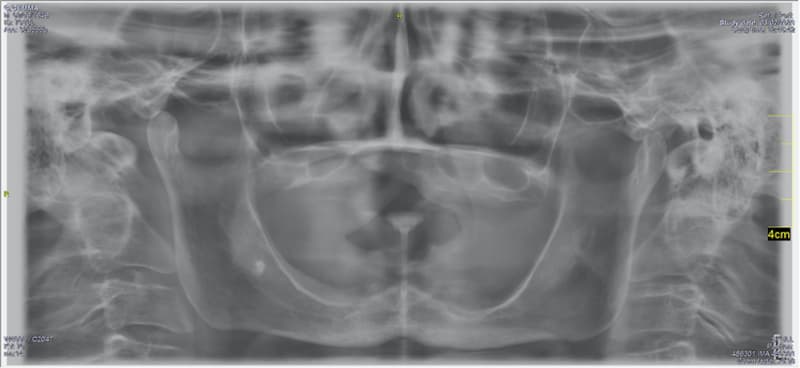
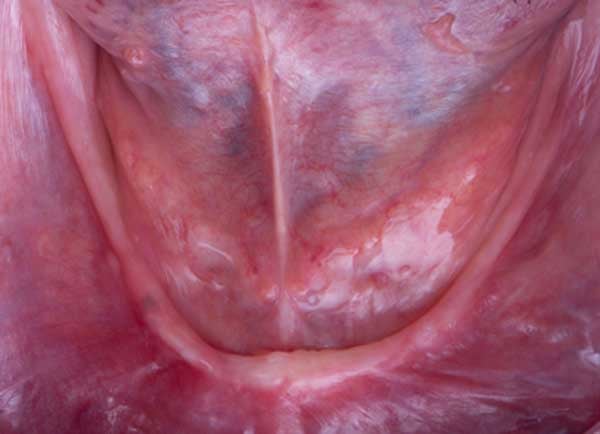
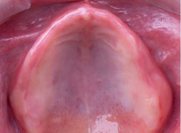
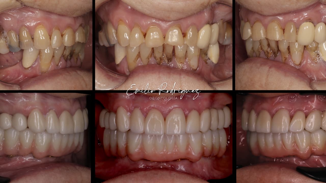
The patient was asked for her medical and dental history and a comprehensive clinical (extra-oral, intraoral, functional, esthetic) and radiologic (panoramic radiograph) examinations were performed. Standardized extra-oral and intraoral photographs of the initial situation with her old removable dentures were taken. Her old dentures were rebased, and a digital impression was taken extra-orally of the rebased dentures. From the available treatment options presented to the patient, she decided to proceed with a double All-On-4: the four zygomatic implant supported fixed prosthesis for the upper and four implant hybrid fixed prostheses for the lower.
The first step of the treatment was to fabricate a new pair of provisional removable dentures with a new intermaxillary relationship, new shapes with aesthetic parameters. The patient accepted the new dentures aesthetically and functionally, and a cone beam computed tomography was performed.
After the planning procedure, the manufacturing of the surgical guide takes place for the upper and lower surgery.


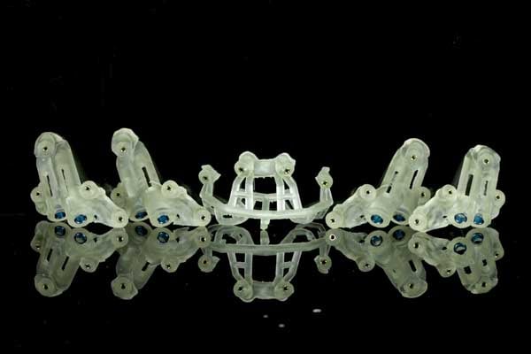
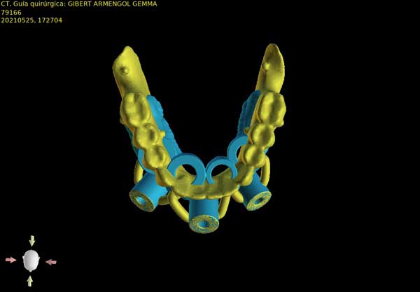
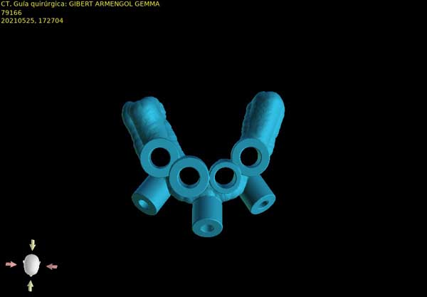
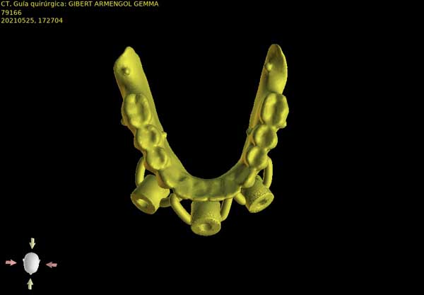
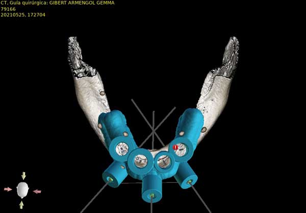

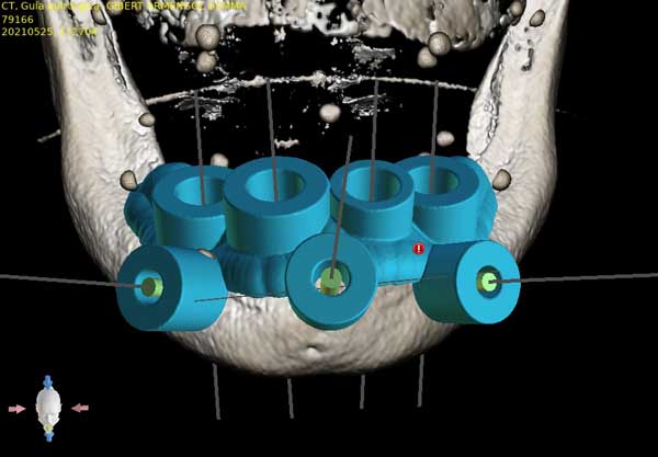
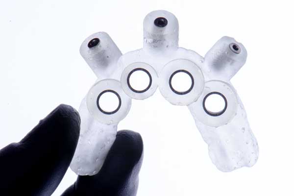

On the upper jaw, the four zygomatic implants were placed guided with the use of the surgical guides and immediate loading was performed with a prosthesis that was produced based on the planning and a guide that reassured the correct position of the loading.
On the lower jaw, the four implants were placed on the planned positions, but one of them didn’t achieve initial stability (10N), so immediate loading couldn’t be performed.
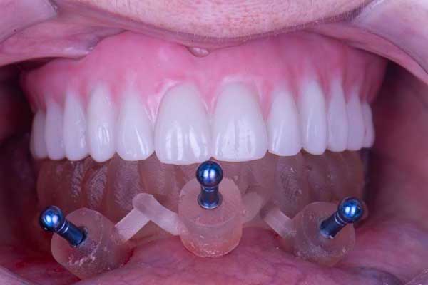
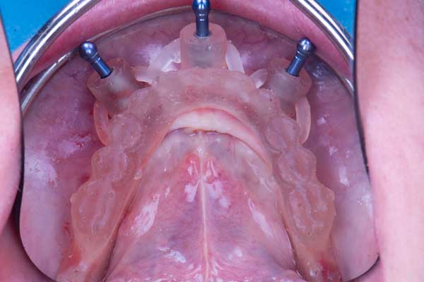
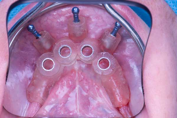
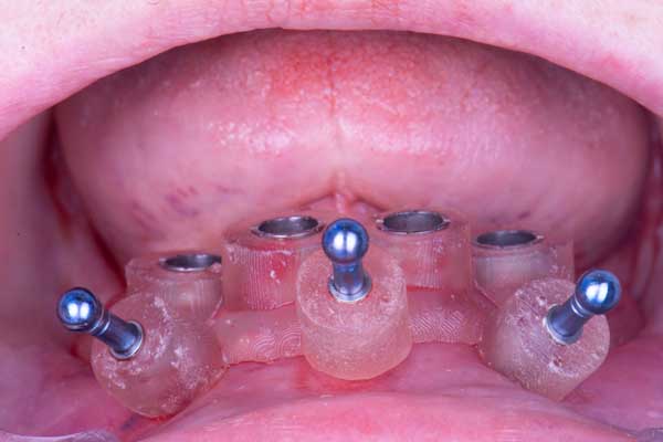
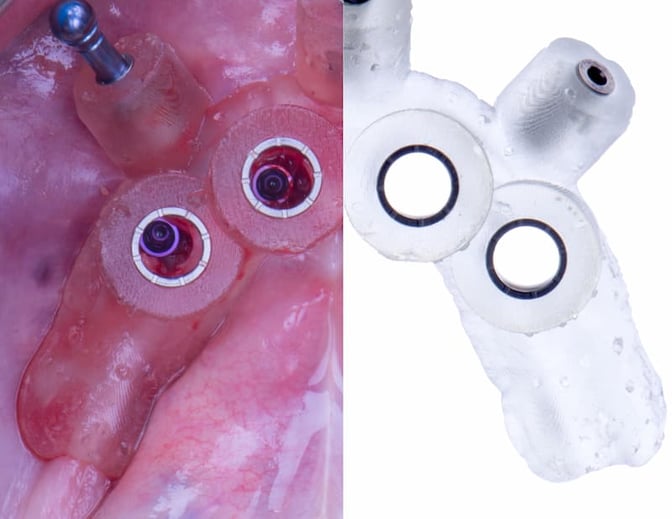
After 3 months, the implant positions of the lower arch were captured with the PIC system. A Medit® intraoral scanner was used to scan the intaglio of the lower denture (once it was rebased with light silicone), as well as the upper and the occlusion.
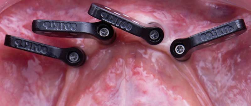
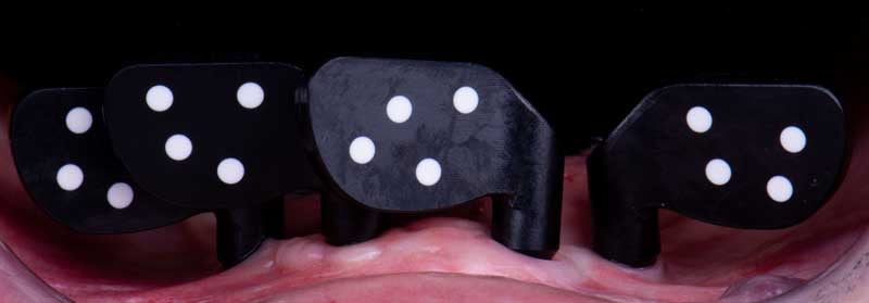
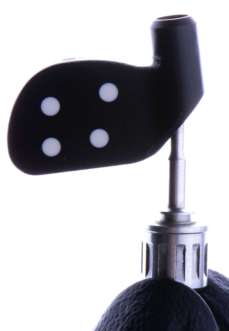
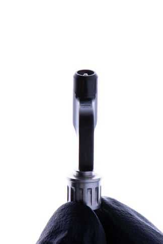
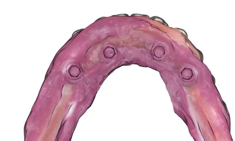
Patient records including the PIC file were then imported in exocad® and the transitional prosthesis was designed and printed in less than 3 hours after taking the PIC system capture.
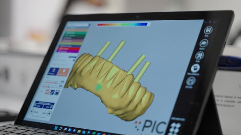 Intaglio design adjustments in exocad®.
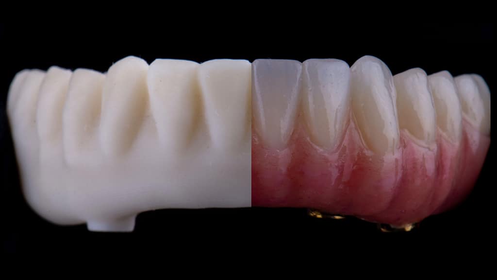
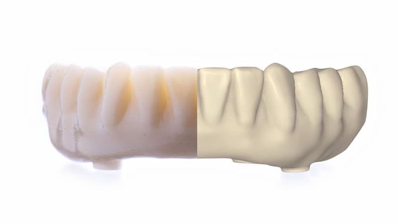 The printed provisional prothesis side-by-side with the digital design.
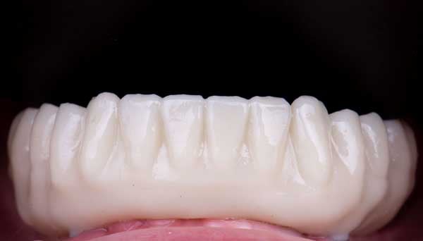
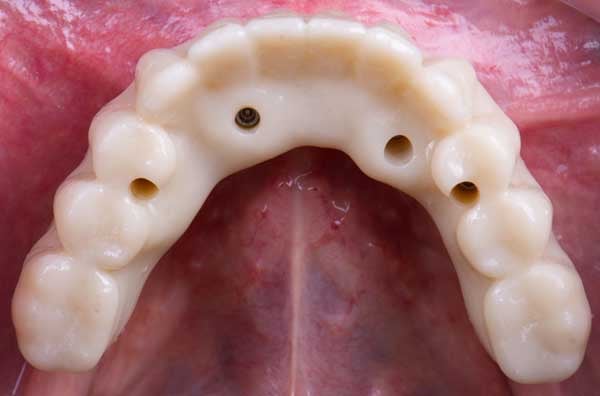
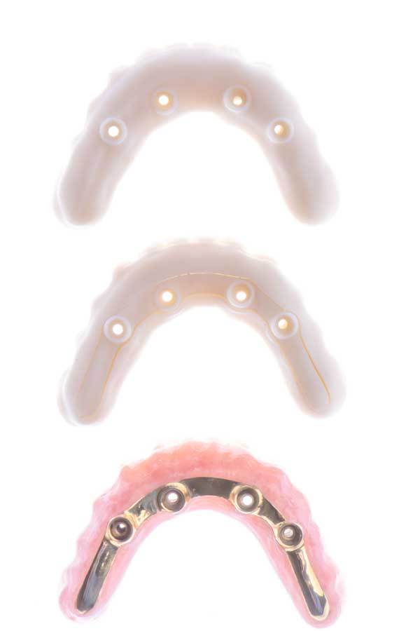
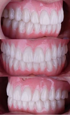
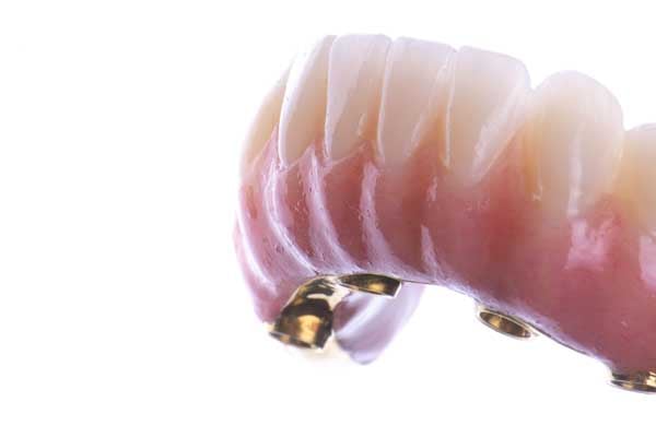
After the evaluation of aesthetics and function for both provisionals of upper and lower jaw during a period of two months, the laboratory then proceeded with the production of both final prostheses directly. The final lower hybrid prosthesis consists of a milled Ti bar and a milled PMMA structure that was cemented on the bar and the upper of a milled monolithic zirconia.
Dental laboratory services were provided by Juan Golobart at Odontecnic (Grupo Ytrio) using Blenderfordental® as the solution to extract the primary bar from the transitional prosthesis to design the final two-part prosthesis.
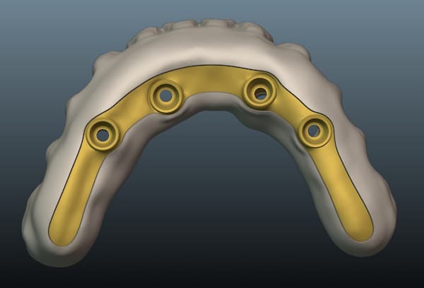
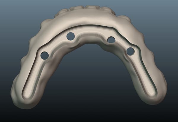
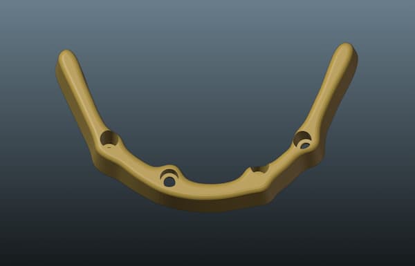
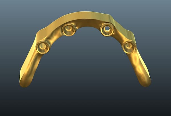
The advantage of digital dentistry is the capacity for an exact “Copy & Paste” of a prosthesis design. In this clinical case this was done twice: first to transition from the provisional removable dentures to the screw-retained transitional prosthesis, and then to produce the final restoration once the transitional prosthesis has been tested clinically.
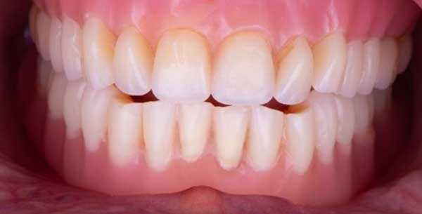
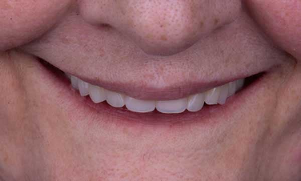
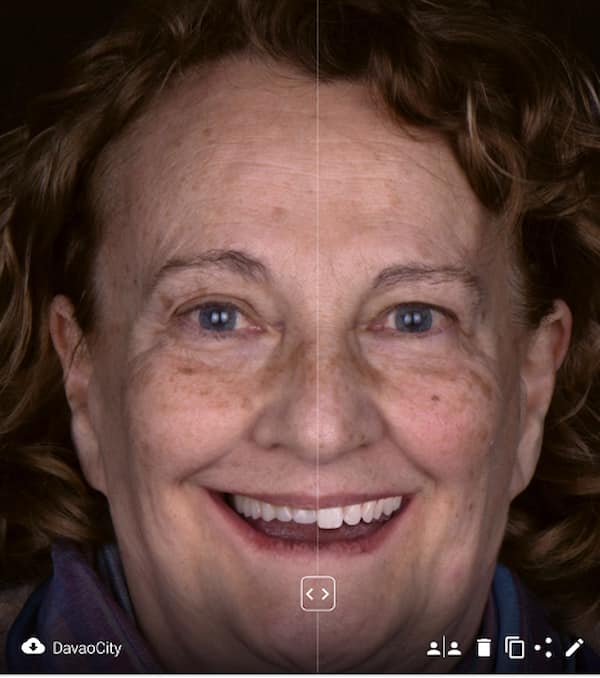
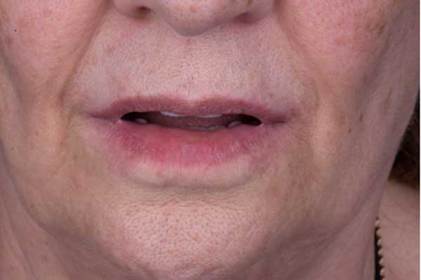
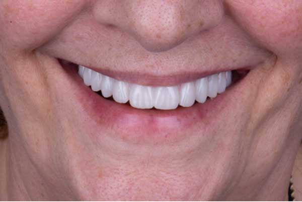
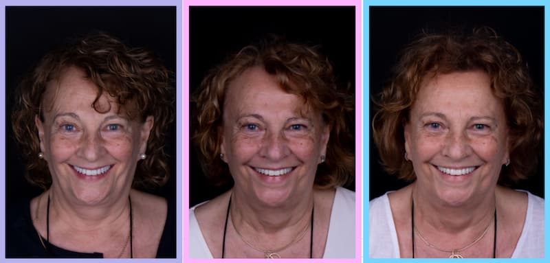 The first extra oral photo is showing the patient wearing the provisional removable denture, the second one with the provisional implant-supported prosthesis and the third with the final restoration.
At the end of the treatment, an occlusal splint was given to the patient to protect from night bruxism.
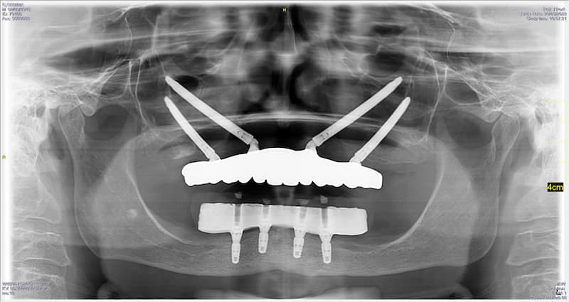
On the radiological analysis, it is shown the perfect adaptation of both prosthesis.

This bimaxillary double All-On-4 case was done by Yolanda Liaropoulou at Universitat Internacional de Catalunya in Barcelona, Spain.
