This video presents an animated overview of the clinical case described on this page (see video on YouTube)
Introduction
Dr. David Lara is a specialist in oral surgery and implantology. In this clinical case, he used PIC system for a maxillary rehabilitation with full extraction of remaining teeth to give the patient the needed corrective treatment.
Clinical case
A 52-year-old male patient, non-smoker and without significant systemic conditions, presented to the Lara & Ochoa dental clinic after experiencing complications from previous dental procedures performed elsewhere.
A thorough intraoral and extraoral examination was performed, complemented by diagnostic imaging, including orthopantomography, CBCT, and intraoral scanning. Clinical findings included:
- Missing teeth: 15, 16, 12, 11, and 25
- Attachment loss on teeth 17 and 27
- Periapical granuloma on tooth 22
- Class III malocclusion
Given the compromised condition of the maxillary dentition and the patient’s esthetic and functional concerns, they proposed a full-arch extraction and implant-supported fixed prosthesis for both arches. The patient opted to proceed with the maxillary arch rehabilitation first.
Initial treatment included basic periodontal therapy to improve periodontal health, consisting of prophylaxis and maintenance.
Following the initial periodontal therapy, Dr. Lara proceeded with a comprehensive digital planning phase. Using CBCT imaging and intraoral scans, they designed a precise treatment plan for implant placement and immediate provisionalization. The strategy involved placing six implants using a post-extraction protocol of all remaining maxillary teeth, combined with bone regeneration techniques using PRGF – ENDORET®. Additionally, soft tissue grafting was planned in the anterior region to restore gingival volume and achieve optimal esthetic contours.
![]()
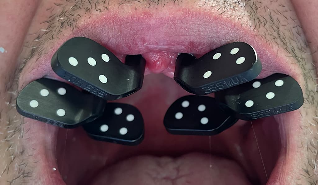 To ensure the highest level of accuracy, implant positions were captured using the PIC system with PIC transfers in an open-flap approach. After completing the capture, healing caps were positioned, and the soft tissues were sutured and scanned digitally to finalize the patient’s master model. This data was then sent to the dental laboratory.
A screw-retained PMMA provisional was then delivered, ensuring proper occlusion and esthetics during the healing phase.
After a four-month osseointegration period, they advanced to the definitive prosthetic phase. Transepithelial abutments were torqued, and a new implant position capture was performed using PIC system to guarantee precision for the final restoration. The process included a metal framework try-in to confirm passive fit and occlusion, followed by a biscuit bake try-in for esthetic and functional validation.
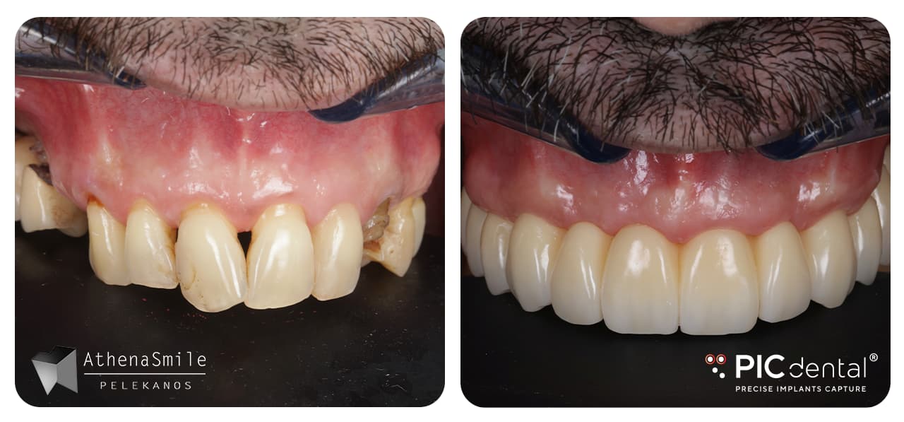
Finally, the definitive screw-retained metal-ceramic prosthesis was delivered, achieving a perfect passive fit and restoring full function and aesthetics.


This case was performed by Dr. David Lara at Lara & Ochoa dental clinic in Logroño, Spain, in 2024.