Inside /PIC_SYSTEM_H_LOGO.svg)
 PIC file
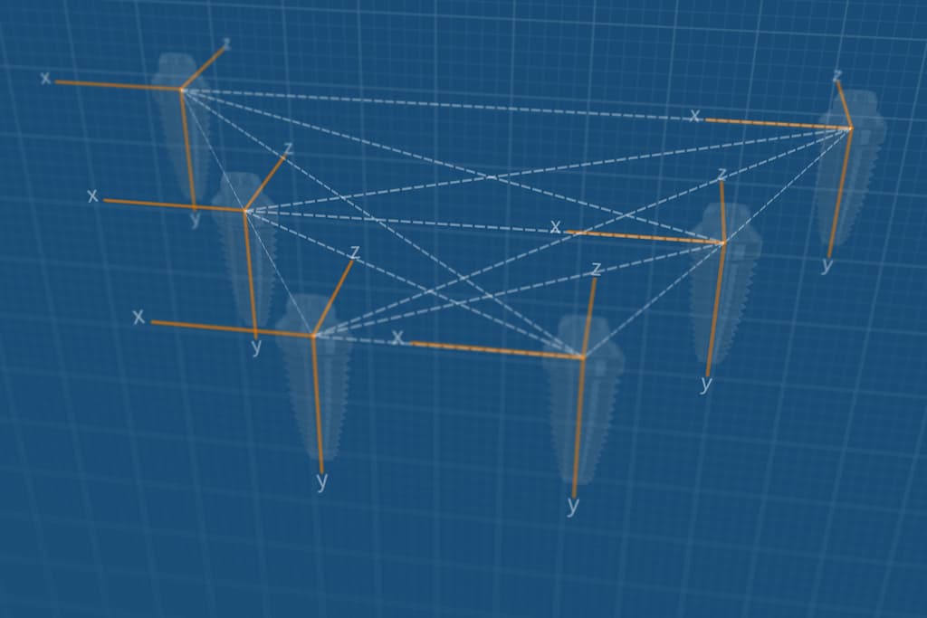
The digital, undivisible vector file containing the implants positions and angulations as measured by the PIC system.
![]() Exported to open STL format
Exported to open STL format
![]() Interrelated implant positions, unchangeable and indivisible after capture
Interrelated implant positions, unchangeable and indivisible after capture
![]() Compatible with all major dental CAD software solutions
Compatible with all major dental CAD software solutions
1. PIC file

Measured by the PIC system
Information: implant positions and angulations
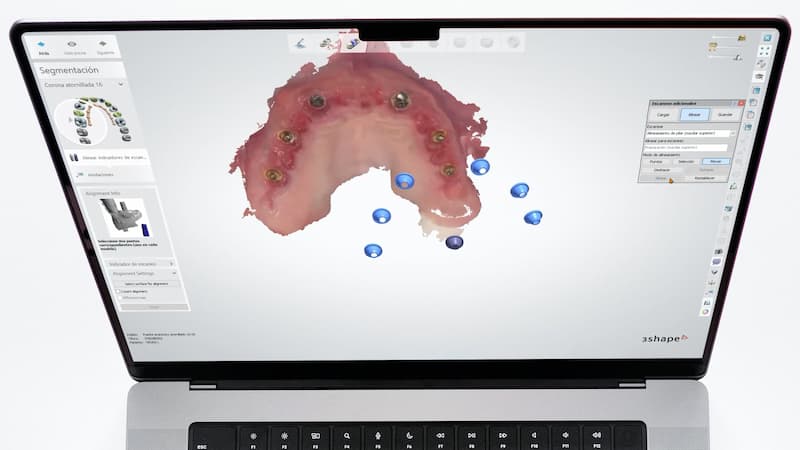
Scan markers: digital (from library)
Export format: STL (scan marker geometry)
Export format: STL (scan marker geometry)

2. Soft tissues
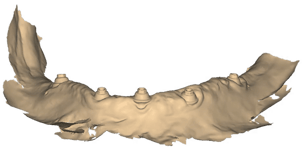
Scanned with an intraoral scanner
Information: soft tissue shape
Scan markers: physical (on patient), scanned
Export format: STL or PLY


/exocad%20logo.png?width=150&height=66&name=exocad%20logo.png)
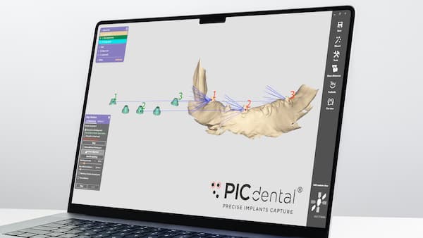
PIC file
How is a PIC file aligned
in dental CAD software?
/exocad%20logo.png?width=200&height=88&name=exocad%20logo.png)